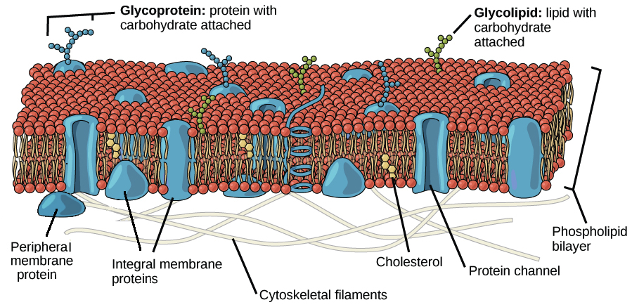
Identify the Plasma Membrane Proteins in the Figure.
The structure and function of the most abundant structural protein of SARS-CoV-2 the membrane M glycoprotein is not fully understood. Cholesterol is a hydrophobic molecule and is found among the hydrophobic tails which you can see in the figure below A critical feature of the plasma membrane is that it is selectively permeable.

Honors Biology Lawrenceville Cells Plasma Membrane Cell Membrane Cell Membrane Coloring Worksheet
The first approach is to identify the receptors by biochemical purification of cellular proteins on the cell surface that bind to the viral antireceptors ie viral structural proteins.
. As a result blood has a higher colloidal concentration and lower water concentration than tissue fluid. For example membrane proteins have hydrophilic and hydrophobic regions that are found among the hydrophilic and hydrophobic portions of the plasma membrane respectively. CellMask Plasma Membrane Stains allow fast and uniform labeling of the cell membrane without the cell-type differences exhibited by lectins.
Proteins enter the Golgi on the side facing the ER cis side and exit on the opposite side of the stack facing the plasma membrane of the cell trans side. It therefore attracts water. New home of TMHMM-20 is.
We previously identified 50 AML-specific plasma membrane PM proteins and 7 of these CD82 CD97 FLT3 IL1RAP TIM3 CD25 and CD123 were implemented in routine diagnostics in patients with AML n 256 and. The plasma proteins suspended in blood cannot move across the semipermeable capillary cell membrane and so they remain in the plasma. Using in silico analyses we determined the structure and potential.
We can also say that the BCOP is higher than the interstitial fluid colloidal osmotic pressure IFCOP. Affinity purification of plasma membrane proteins using the viral structural proteins as a ligand is feasible. The structural proteins of SARS include membrane glycoprotein M envelope protein E nucleocapsid protein N and the spike protein S.
Differences in signaling and drug sensitivity of such subclones complicate treatment and warrant tools to identify them and track disease progression. Alternatively immunoprecipitation of plasma membrane proteins that bind to the viral. CellMask plasma membrane stains may be used for translocation assays plasma membrane dynamics and cell segmentation tool for high-content screening as well as to stain cellular plasma membranes for standard fluorescence.

Unit 1 Practice Questions 60 Questions Flashcards Quizlet

Membrane Protein Overview Creative Biolabs Blog
Structure Of The Plasma Membrane Article Khan Academy

Ch 4 Quiz Flashcards Quizlet

Plasma Membrane Definition And Examples Biology Online Dictionary

Mastering A P Chapter 3 Cells The Living Units Diagram Quizlet

Mastering A P Chapter 3 Cells The Living Units Diagram Quizlet

Structure Of The Plasma Membrane Article Khan Academy
The Extracellular Matrix And Cell Wall Article Khan Academy

Membrane Proteins Advanced Read Biology Ck 12 Foundation

Difference Between Channel And Carrier Proteins Difference Between
2 6 Membrane Proteins Biology Libretexts

Mastering Bio Chpt 7 Flashcards Quizlet

What Is The Difference Between Integral Peripheral And Surface Proteins Compare The Difference Between Similar Terms

Pin On Physiology

Membrane Proteins Ck 12 Foundation

Difference Between Intrinsic And Extrinsic Proteins Compare The Difference Between Similar Terms

Difference Between Cell Membrane And Plasma Membrane Definition Composition Function

Mastering A P Ch 03 Hw Diagram Quizlet
Comments
Post a Comment